One of the main challenges for science and engineering education is to captivate the enthusiasm of students, especially during their early college experience. Computer-Integrated Surgery is especially difficult to teach because the subject is often overwhelming in its complexity and strong math and scientific computing skills are needed for even the simplest hands-on project. The challenge of such a course is to strike the right balance between presenting captivating technologies and assignments.
- Introduce students to practical biomedical engineering applications. In particular, provide experience in computer-assisted surgery in affordable and reproducible lab environment.
- Enhance understanding of concepts learned in class through hands-on practical experience.
- Attract students to advanced studies in biomedical technology.
Background
 One of the main challenges for science and engineering education is to captivate the enthusiasm of students, especially during their early college experience. Computer-Integrated Surgery is especially difficult to teach because the subject is often overwhelming in its complexity and strong math and scientific computing skills are needed for even the simplest hands-on project. The challenge of such a course is to strike the right balance between presenting captivating technologies and assignments.
Dr. Fichtinger, teaches Computer-Assisted Integrated Surgery at Queen’s at three levels, tailored to various cohorts: graduates, senior undergrads, and junior undergrads. These courses are designed to introduce students to the concepts and issues of Computer-Integrated Surgery. The students learn to ask questions and look for answers in the same way clinical engineers analyze and build systems. Multi-disciplinary concepts and systems are emphasized through a series of novel clinical applications that are currently in use or under development at various institutions, primarily by biomedical computing faculty at Queen’s. The homework assignments pertain to medical image processing, surgical planning, surgical navigation and medical robotics. While the assignments across different classes tend to cover similar problems, the math and programming skills required are tailored to the particular audience. For example, the freshmen assignments do not require programming skills. At more senior levels, however, the complexity of problems increases to resemble real life surgeries.
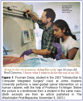
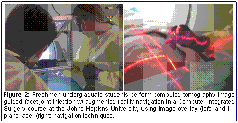
 Dr. Fichtinger taught Computer-Integrated Surgery courses at the Johns Hopkins University in the United States. The course was featured in the Washington Post Magazine (Figure 1, full article is attached.) Between 2003 and 2007, the course included a one-week clinical laboratory module that included human cadaver surgery (Figure 2). The human cadaver surgery module was the pinnacle of the course, making an impact on the career choice of many students in the past. This fact is evidenced by student testimonials, available in the above mentioned Washington Post article, and through course evaluations.
Teaching a human cadaver surgery lab, however successful it has been, is not readily adoptable by other biomedical faculty and institutions for many reasons: the timely availability of human cadavers tends to be generally difficult; special facilities, such as operating room and clinical quality medical imaging devices needed; ethics board approval, covering all stages and aspects of the cadaver surgery is required; participating students must have the highest level biohazard blood-born pathogen safety credentials; some students may reject human cadaver experiments for religious reasons; and, teaching human cadaver surgery requires surgical skills and qualifications from the instructor and staff. These reasons prompted us to propose a universally accessible and transferable method for teaching computer integrated surgery.
The Perk Station
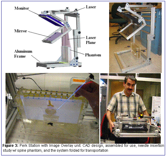
The Perk Station is an inexpensive, simple and easily replicated surgical navigation workstation for hands-on practice with computer assisted surgery on non-biohazardous specimens in a standard ‘dry’ laboratory. (Figure 3).

Dr. Fichtinger’s research team has previously developed state of the art virtual reality surgical navigation techniques for percutaneous (through the skin) interventions. Techniques include image overlay [1] and tri-plane laser overlay [2] (see Figure 2). We re-engineered these clinical systems for laboratory use, by taking away complex features that were required for patient safety and clinical compatibility. We comprised the remaining functions in the Perk Station, a stand-alone laboratory workstation for percutaneous surgery. The Perk Station allows students to plan and perform computed tomography and magnetic resonance image guided needle based surgeries under virtual reality navigation. Students plan the surgery through computer interface, by calculating target locations, tool trajectories and other critical parameters. They will perform the planned intervention on a phantom, an anatomically realistic artificial object. Finally, they evaluate their own performance relative to the surgical plan. The Perk Station supports a variety of interventions, chiefly depending on the tuning of the surgical planning interface software and the phantom. We start with spinal interventions and thermal ablation of solid tumors, such as liver cancer. Phantoms have been fabricated from multiple layers of tissue-equivalent transparent gel in a clear acrylic container. During the training surgery, we drape the phantom and prep the site, just as in actual clinical use. Upon completion of the intervention, we uncover the phantom and visually assess the accuracy of the procedure and estimate the extent of collateral tissue damage and morbidity.
Applicability
The Perk Station is easily be replicable and adaptable for teaching computer assisted surgery at all levels, from graduate courses to high-school science classes. The Perk Station allows the instructor to demonstrate different technical and medical aspects of complex biomedical engineering systems. Specific disciplines represented in the Perk Station include computing, mechanical engineering, electrical engineering, and several disciplines of modern medicine and biology, such as medical imaging, minimally invasive surgery and physiology. Hence a wide range of audience can connect with the Perk Station, from unassuming high school students to seasoned clinicians and medical engineering experts.
The Perk Station is portable and fits in a suitcase when disassembled. In addition to courses, the Perk Station can also be used in demonstrations at various events, such as student fairs and open houses. It is also be an ideal demonstration tool for the Computing High-Tech Academic Mentorship Program (CHAMP) in their regular nationwide high school presentation tours. Due to its reproducibility and easy access, it can fulfill an important role in reaching out to historically under-represented minorities.
The apparent simplicity of the Perk Station should not belie its potentials in teaching and training medical professionals, particularly medical students and residents. There is a general misperception and under-appreciation among the public of the skills required for needle based surgeries. In reality, trainees gravitate to learning centers where procedural skills are taught. There is also popular trend to minimize the steep learning curve by using simulations. Patients and patient advocates are less tolerant of training on clinical cases. Increased clinical workloads have also demanded increased provider productivity. The changing financial climate and commercial initiatives have catapulted to the forefront the need of training and performance evaluation without involvement of human subjects. Static or declining reimbursements have driven the need for economical solutions: training systems of with accuracy, efficiency, simplicity, and low cost. The Perk Station fits in this trend eminently.
Transferability
The final design of the Perk Station, including hardware blueprints, phantoms blueprints, and software source code will be made publicly available trough the National Alliance for Medical Image Computing (www.na-mic.org). Simple design and low costs allow all interested parties to replicate the hardware and install the software. The Perk Station is modular, so users could further downscale its functions and thus save on hardware. The distribution website will also supply medical image data and pre-made surgical plans so that users can operate the Perk Station without having access to medical imaging facilities. There will be appropriate documentation and tutorials provided on the website. It most be pointed out that a significant fraction of our costs include design costs: solid models, blue prints, CNC machine programs, laser cutter settings, etc. As the complete design will be available free of charge, actual replication costs will be significantly less then our current budget. Multiple replicas will further cut the costs, nearly to costs of raw materials.


